Ventral Slot Technique
Gingi McLeod Age: 8 years Breed: Rottweiler Sex: F/S
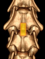
A ventral slot consists of a partial corpectomy of two adjacent vertebral bodies and the corresponding intervertebral disc to access the ventral vertebral canal.1 Laryngeal paralysis (LP) is a common respiratory disorder that implicates loss of normal function of the larynx where the arytenoid cartilages and consequently the vocal folds remain.

Ventral Slot Decompression: This is a surgical procedure that is performed to remove herniated intervertebral disc material from the cervical region of the vertebral column. Ventriculoperitoneal Shunt Placement: This is a surgical procedure where excessive fluid pressure within the central nervous system is relieved by placing a sterile stent. The standard ventral slot had a significant higher increase in ROM in extension compared with the other three techniques. The slanted slot had a significant lower increase in ROM in flexion. Discussion/Conclusion The described ventral slot techniques have similar biomechanical effects on the canine cervical vertebral column.
At 7 months of age, Gingi was adopted by the McLeod’s from a local shelter and had been an otherwise healthy house pet. Around 11 months ago, Gingi would intermittently cry out in pain after performing certain activities, such as climbing stairs or getting into or out of the car. Gingi also started exhibiting signs of ataxia and rear limb weakness as her signs progressed.
Gingi was referred to Bush Veterinary Neurology Service where an MRI was performed. The results of the MRI showed Gingi had a condition termed Wobbler’s Disease, which resulted in cervical disc protrusions at C4-5 and C5-6. Gingi underwent a surgery called a ventral slot at C5-6 with a fenestration at C4-5. Following surgery, Gingi was sent home on strict bed rest. The McLeod’s were to perform passive range of motion exercises on all 4 limbs and to assist her into a standing position and to go outside with the aid of a help em up harness or sling a few times daily. In her initial post-op period Gingi was not able to urinate on her own and was given medication to help with bladder expression. Gingi also received hyperbaric oxygen treatments several times in the month following her surgery.
Gingi presented to Skylos Sports Medicine to start rehab therapy within a few weeks of surgery. At this time Gingi was too weak to support herself in a standing position, but would move all 4 legs in an attempt to walk with assistance. Her owners were performing passive range of motion exercises 2-4 times daily and assisting her to stand for short periods of time. They felt Gingi had some awareness of her bladder and rectum as she would often vocalize when she had to urinate or defecate. Gingi mainly lied on her side, but was able to get herself into a sternal position if she was lying on her left side. She was unable to accomplish this on her right side. She had anal tone and was able to wag her tail. Gingi had delayed withdrawal reflexes in all 4 legs, but was able to move her legs if persuaded with treats.
At Gingi’s first rehab session we spent time getting to know her and assess what would be good ways to assist her recovery that worked with her personality and with what her owners could accomplish at home as well. Therapy for her first session included; massage and manual techniques, non-thermal class IV laser and specific exercises to promote leg movement and proprioception. One such exercise involved assisting Gingi to drape her body up and over an exercise peanut and rolling her back and forth so both her front paws and rear paws would touch the floor. This is a good exercise to promote proprioception as well as support her body while stabilizing muscles engage.

The McLeod’s were sent home with various exercises, bodywork instructions, and ongoing care parameters. It required daily dedication on her owners part to provide passive range of motion, sensory work such as brushing her in specific ways and helping her body and limbs re-pattern things like sitting up and the steps it takes to accomplish this. Multiple short sessions of standing and assisted walking were also assigned. In addition, Gingi visited Skylos for rehab twice weekly.
Week 1: Gingi had made a few small improvements. Her withdrawal reflexes were stronger in all 4 limbs, she was able to remain sternal on her right side for a few moments and she attempted to stand for a bully stick. However, Gingi was too weak to support her weight by herself when trying to stand. Therapies included; massage, laser and exercises to promote leg movement, balance and proprioception.
Week 2: Gingi continued to make improvements. When taken outside, she was able to stand for a few moments and walked a small distance with little assistance. At this time, her rear legs had more strength and when she tired, Gingi tended to collapse on her front legs. As Gingi would tire rather quickly, we would exercise for a small period of time and then rest. During the second session of the week, Gingi was able to remain in a sternal position on her right side without assistance. Gingi was now also being treated for a UTI. Once a negative urine culture was obtained, the plan was to start Gingi in the underwater treadmill. Therapies included; massage, laser and exercises to promote leg movement, strength, balance and proprioception. The McLeod’s continued a lot of home exercises and mainly concentrated an assisted standing and walking.
Week 3: I was really impressed with Gingi’s improvements. The owners were doing a wonderful job with her home exercises. Gingi was able to walk around with little assistance for around one minute before she tired and tended to collapse. She was also able to stand on her own with little assistance for 30-45 seconds. Gingi was strong enough to walk over 2 inch cavaletti rails for 3 passes. Therapies included; massage, laser and exercises to promote strength, balance and proprioception. Gingi’s urine culture came back negative, so for her second session of the week, she tried the underwater treadmill. Gingi walked for a total of 6 minutes with rests every 2 minutes. She would occasionally knuckle on the left front leg, but overall, did very well.

Week 4: Gingi continued to gain strength at home and was now strong enough to potty on her own outside and no longer needed assistance with bladder expression. Another step in a positive direction that Gingi made at this time was that she was now able to stand on her own from a sit or down position with little to no assistance. She still tired rather quickly, so we would exercise in small increments. Therapies included; massage, laser, underwater treadmill and other exercises to promote strength, balance and proprioception, such as cavaletti rails, figure 8’s and assisted sit to stands.
Week 5: During this week, Gingi had a re-check appointment with the neurologist and she was very happy with Gingi’s progress. During her sessions, Gingi was able to walk around without any assistance and even walked over the 2 inch cavaletti rails on her own. Therapies included; massage, laser, underwater treadmill, assisted sit to stands, cavaletti rails and figure 8’s. Her owners continued to diligently work with her at home.
Weeks 6 and 7: Gingi developed another UTI, so she was not able to go in the underwater treadmill. Her owners felt she was a bit weaker in the rear legs, but overall, was walking well. Gingi had an ataxic gait, but was able to walk around without any assistance. Gingi’s conscious proprioception was slightly delayed, but she was able to “right” all of her paws. Therapies included; massage, laser, sit to stands and started the wobble board and balancing on proprio discs.
Weeks 8 and 9: Gingi continued to do well. She would occasionally knuckle on her left front leg, but this had always appeared to be her weakest leg. This week, Gingi decided she no longer cared for the underwater treadmill and refused to walk in it. During this time, she became able to retrieve treats off of the floor without stumbling or falling. Therapies included; massage, laser and exercises to promote strength, stamina and balance. Her rehab sessions were decreased to once weekly.
Weeks 10 and 11: Gingi continued to recover her strength and was beginning to gain muscle mass. Her gait was less ataxic and she was now able to trot. Gingi’s conscious proprioception continued to improve. Therapies included; massage, laser and exercises to promote strength, stamina and balance. She was doing well her sessions were decreased to every other week.
Gingi comes twice a month for rehab therapy. She is doing well and our work is to continue building on her strength and stamina. Gingi has gained in energy, but still tires after 15 minutes of work. She has been an absolute joy to work with. Gingi has a strong will and was quite determined to walk again. Her owners are also lovely people and were dedicated to her recovery.

Laura Martino-Calfo, RVT,CCRP,CCMP Dr. Faith Lotsikas, CCRT
Skylos Sports Medicine
OBJECTIVE: To describe the use of a video telescope operating monitor (VITOM™) for ventral slot decompression and to report its clinical applications using preoperative and postoperative computed tomography (CT) myelography.

STUDY DESIGN: Prospective case series.
ANIMALS: Consecutive dogs presented with cervical intervertebral disc disease requiring surgical decompression (n = 30).
METHODS: Demographic data, preoperative neurological status, localization and lateralization of the compression, total operative time, surgical complications, ventral slot size and orientation, hospitalization time, and postoperative outcome were recorded. Preoperative and postoperative spinal cord area at the compression site and ratios of compressed to normal spinal cord area were calculated by CT myelography.
Ventral Slot Technique Meaning
RESULTS: French Bulldogs were the most common breed of dogs (n = 15; 50%) and neck pain was the most common neurological sign (n = 18; 60%). Postoperative CT myelography confirmed that spinal cord decompression, postoperative spinal cord area, and the ratios of compressed to normal spinal cord area improved significantly compared with preoperative measurements (P = .01). Sinus bleeding occurred in 20% of dogs. The mean ratios (± SD) of ventral slot length and width compared with vertebral body length and width were 0.21 ± 0.08 and 0.31 ± 0.07, respectively. The mean postoperative hospitalization time was 3.0 ± 0.6 days and all dogs showed clinical improvement and an excellent outcome.
Ventral Slot Technique Game
CONCLUSION: The VITOM™ ventral slot decompression technique was fast and easy to perform. It allowed a minimally invasive approach with a small ventral slot while improving spinal cord visualization. The results of this study support the use of the VITOM™ technique in spinal veterinary surgery.